Prostate Cancer Diagnosis

Visit to the General Practitioner or Urologist is the first step in the diagnosis process. In the context of urinary tract infection, presence of blood in the urine or sperm, prostatitis, urinary discomfort or urine retention your doctor will perform specific tests to assess irregular consistency of the prostate.
During the medical visit the doctor will ask you about your personal and family background. He or she may also inquire about any signs you might be having, such as urination issues or the appearance of blood in your urine. Then, the doctor will perform a clinical exam*. This implies a digital exam and sometimes an ultrasound exam to assess the size and consistency of the prostate gland, as well as any palpable abnormalities (increase in size, hardened area, etc.). Additional tests may also be recommended by your doctor.
Basic Tests
PSA (Prostate Specific Antigen)

PSA (Prostate Specific Antigen): This test is considered by many physicians as an important diagnostic tool to detect the presence of prostate cancer. When prostate cancer develops, the PSA level usually goes above 4 ng/ml (nano grammes per millilitre), depending on the patient’s age. But it is important to remember that about 15% of men with a PSA below 4 ng/ml (nano grammes per millilitre) will have prostate cancer on biopsy. If your level is in the borderline range between 4 and 10, you have about a 25% chance of having prostate cancer. If it is more than 10, your chance of having prostate cancer is over 50% and increases more as your PSA level increases.
You might also have an elevated PSA for other reasons like prostatitis or benign hyperplasia. If your PSA level is high, your doctor may recommend a prostate biopsy to find out if you have cancer.
Digital Rectal Exam (DRE)
During a DRE, the physician inserts a gloved, lubricated finger into the rectum and examines the prostate for any irregularities in size, shape, and texture. Often, the DRE can be used by urologists to help distinguish between prostate cancer and non-cancerous conditions such as BPH.

Biopsy and rectal ultrasound
Performed under local anaesthesia or sedation, prostate cell samples are collected with a needle, guided by rectal ultrasound. It is an outpatient technique. Sometimes a massive biopsy is required under spinal anesthesia requiring a short hospital stay. Eventually these images can be compared with a previous MRI. Then, after a process that can last a week, these samples are analysed under a microscope by a pathologist to see how irregular the cells are.
The number of positive biopsies (those containing cancer cells), the features of the tumour tissue, and the cancer cells' extension outside the prostate capsule are all evaluated. When biopsies reveal the existence of cancer, further examination can be performed, based on the cancer's characteristics.
Gleason Scores:
The Gleason test is a grading scale that indicates the current aggressiveness of the tumour and helps the physician determine how likely a patient’s cancer may spread. Tissue removed from the prostate during biopsy is examined microscopically and graded. The higher your Gleason score, the more likely it is that your cancer will grow and spread.
Based on your PSA, Gleason score and digital rectal examination, your doctor can prescribe additional tests after you’ve been diagnosed with prostate cancer to learn more about the possible extension of the disease and select the most appropriate treatment for your situation.
Additional tests
Computed Tomography (CT scan):

This is a form of X-ray that provides a clear image of the tissues inside your body. It is used to look for cancer spread in lymph nodes or distant organs. Before the exam, you might be asked to fast for a few hours. To help light up areas of your body on the scan, you might be given an injection or a special drink.
If you're allergic to iodine or have asthma, tell the radiographer before you take the drink. For a few minutes after the injection, you may feel hot all over. You will lie on a table that will move through a huge doughnut-shaped system during the scan. It takes between 10 and 30 minutes and does not hurt.
Magnetic resonance imaging (MRI scan)
This is a scan that uses magnetic fields to create an image of the body's tissues. It allows to see the prostate with great precision and distinguish tumor growths. During the procedure, you might be given an injection to highlight specific areas of the body. Prior to it, your clinician will discuss the procedure with you to make sure you’re not allergic to any of the component that will be injected. You'll be lying inside a tunnel-like machine for the duration of the evaluation. Some people are concerned that they will become claustrophobic. If you're worried, make an appointment with the radiographer the day before. They might be able to prescribe you some medicine to help you relax during the day. Please let you physician know in case you have any medical device in your body. Metal jewellery is not permitted. The scan is painless, but it can be very noisy. Earplugs will be sent to you. It is an outpatient test.
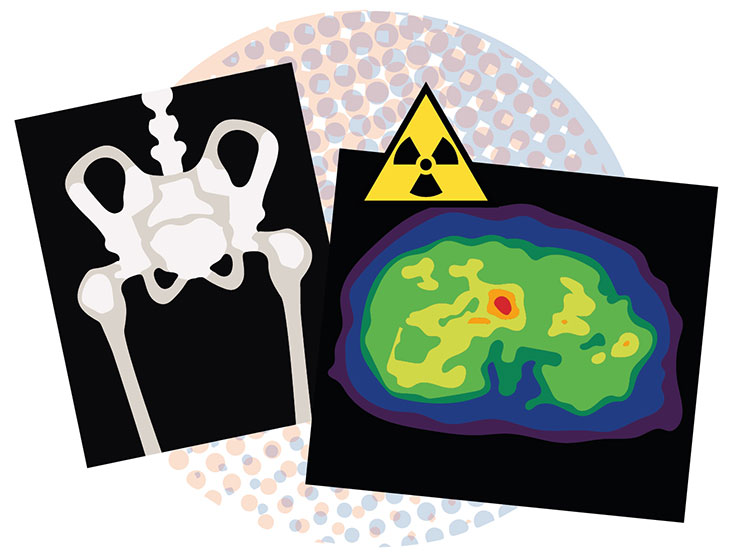
Bone Scan

Bone scans are very sensitive and can identify cancer cells in the bone before an X-ray can detect them. A small amount of mildly radioactive material is inserted into a vein, normally in your arm, for this examination. You must wait up to 3 hours after the injection before the scan can be performed. You might want to bring a book or a friend with you to keep you company. Many of the bones in your body are scanned. Since abnormal bone absorbs more radioactive material than normal bone, it can be detected on a scan. The radioactivity used in these scans is extremely low and completely healthy. It vanishes from the body in a matter of hours.
However, interaction with pregnant women and very near contact with babies or young children should be avoided for at least 24 hours (such as holding them or letting them sit on your lap).
Positron emission tomography (PET scan)
This technology associates a CT-Scan and a metabolic imaging. The metabolic imaging consists in the injection of a radioactive tracer
Contact Support