true
Icon to close the modal
Contact Us
Sales
Modal Close Icon
Icon to close thank you modal
Thank you for contacting our sales team!
A sales representive will get in touch with you shortly.
Customer Care
Phone Icon
0800 917 8776
BDUK_customerservice@bd.com

Phasix™ Mesh  

Composed of material derived from a fermentation process, Poly-4-hydroxybutyrate (P4HB), Phasix™ Mesh provides critical strength during the initial healing phase.

Contact Us
Phasix™ Mesh
Loading

Overview

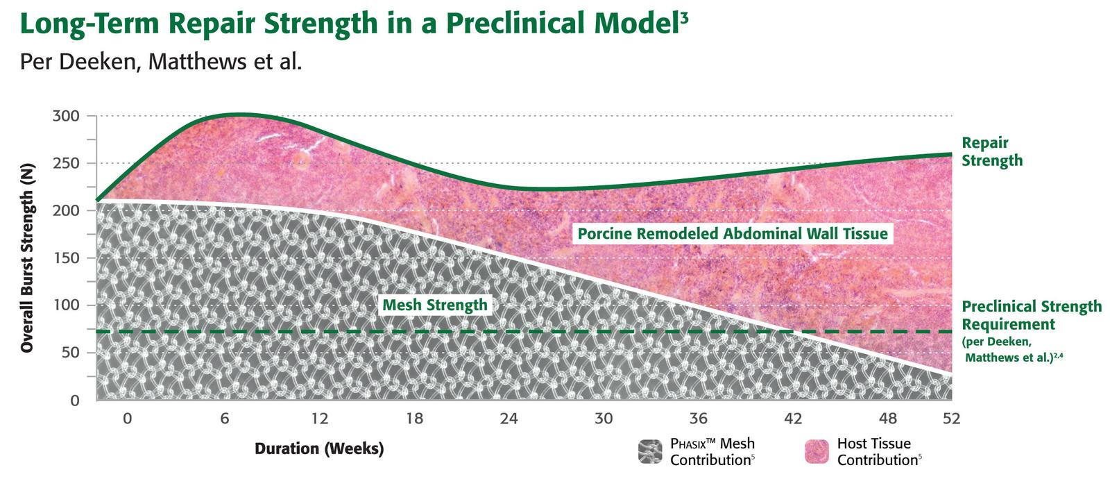
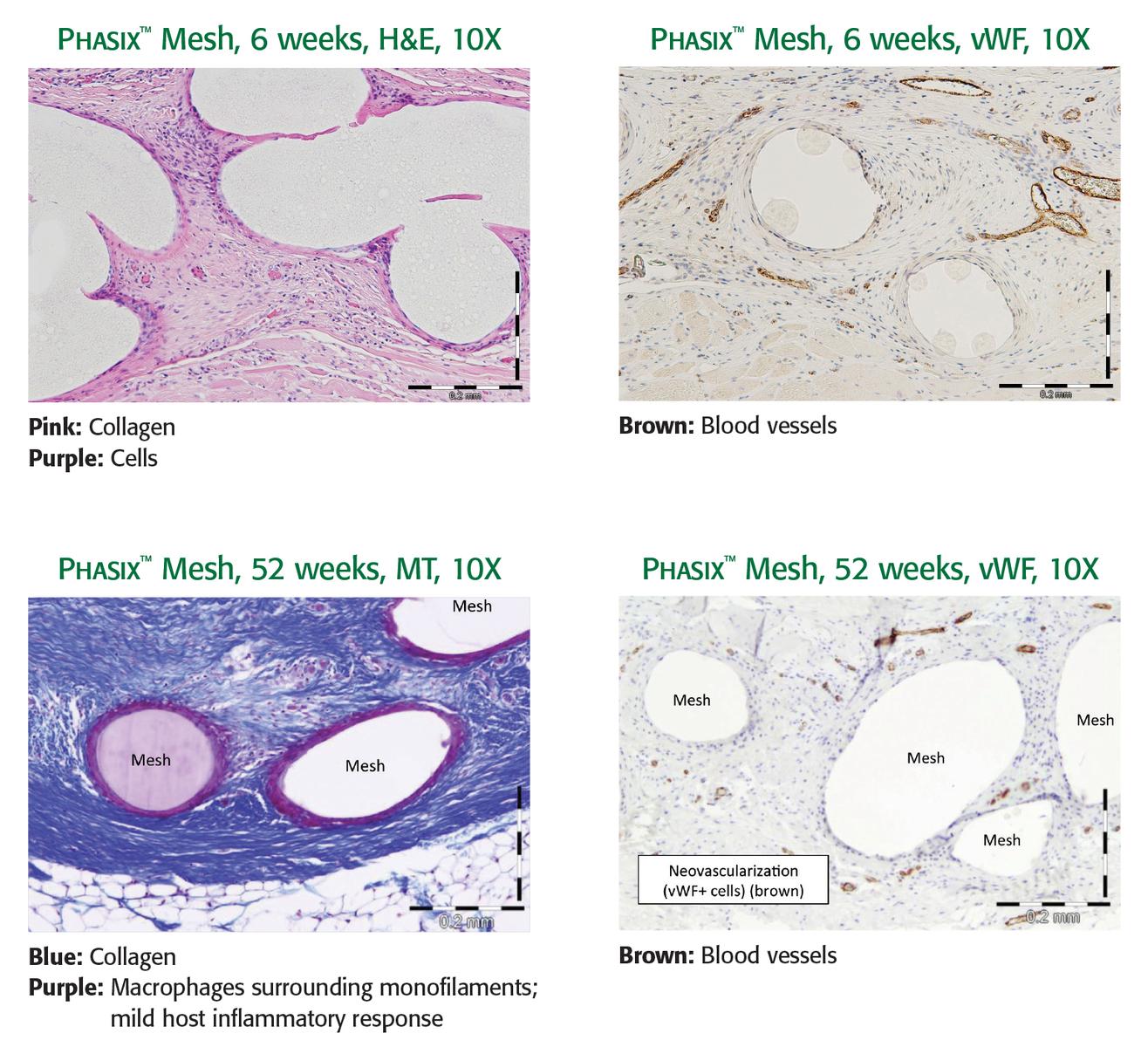
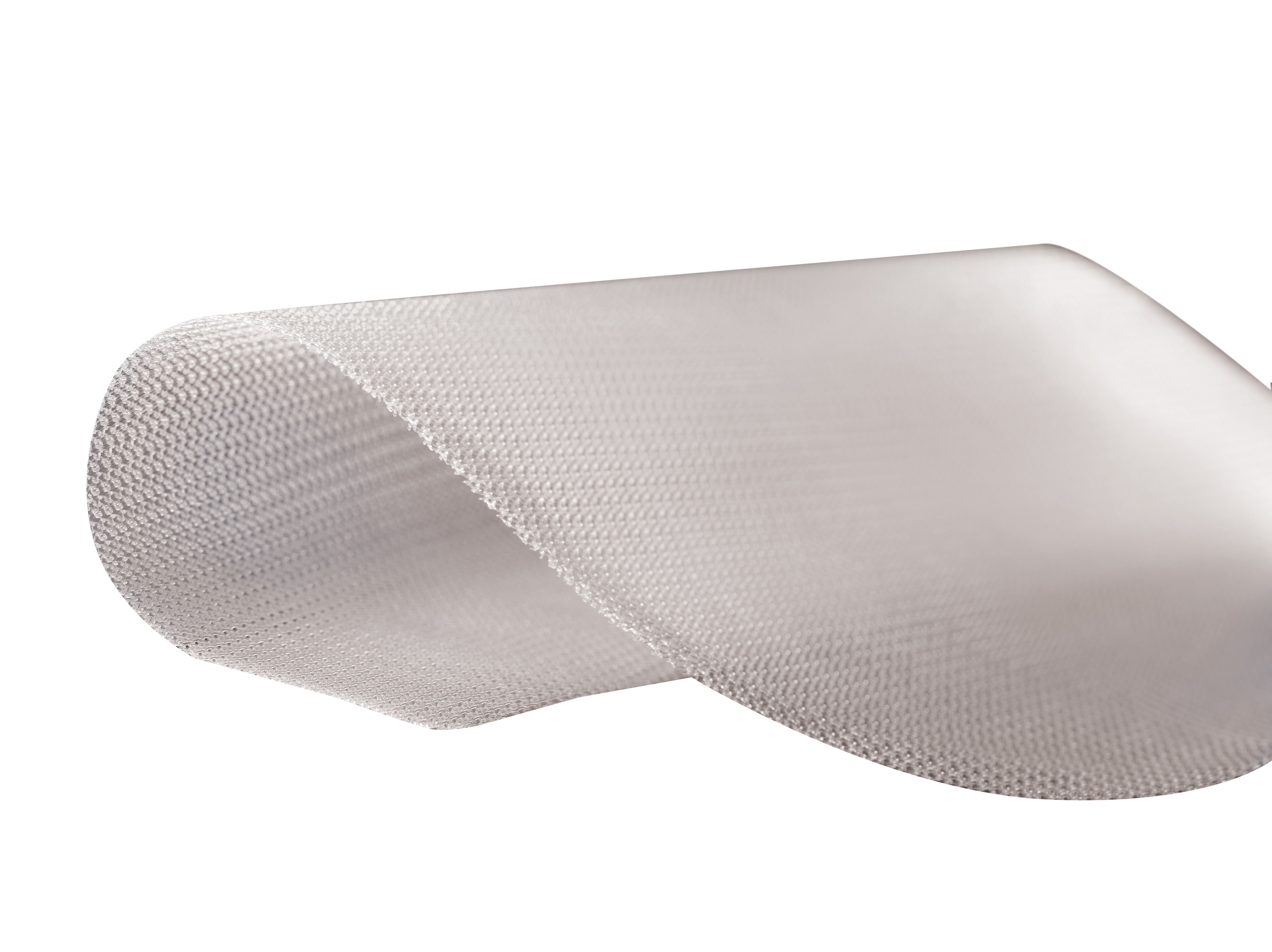
Designed to enable constructive and functional tissue remodeling1

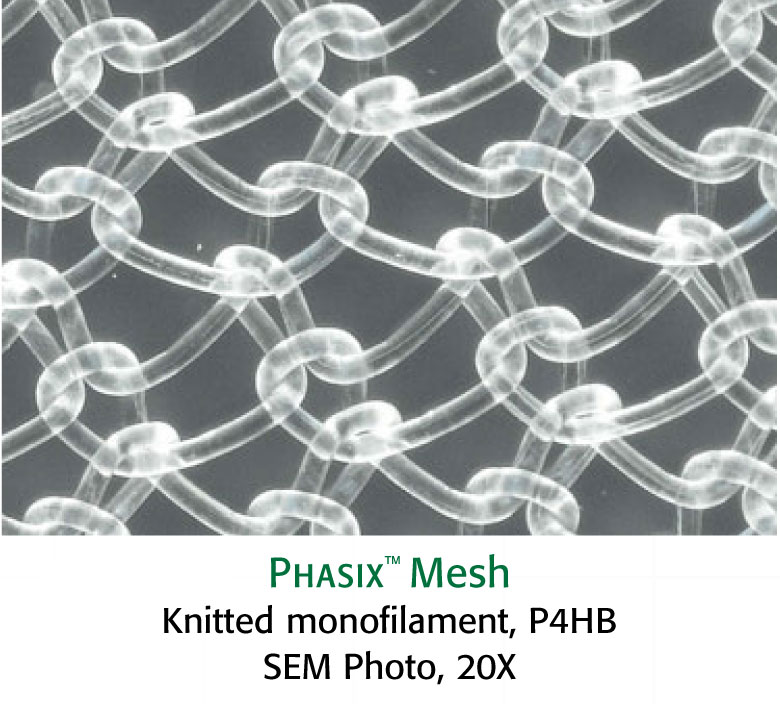
Phasix™ Mesh provides a fully resorbable monofilament scaffold for rapid tissue incorporation that has been designed to allow for the repair strength of a synthetic mesh along with the remodeling characteristics of a biologic graft.1

true
Features and Benefits

THE NEXT PHASE IN HERNIA REPAIR

Repairs1
The open monofilament mesh scaffold provides early integration and repair strength*

Remodels1
Preclinical testing confirms vascular integration and incorporation, with abundant mature collagen at 52 weeks. Gradually transfers load to native tissue over time*

Restores1
As Phasix™ Mesh is remodeled, it is replaced with functional tissue, ultimately resulting in a strong repair at one year*

WHAT IS PHASIX™ MESH?

Phasix™ Mesh is a knitted monofilament mesh scaffold using Poly-4-hydroxybutyrate (P4HB), a biologically derived, fully resorbable material.

  • Monomer form (4HB) is a naturally occurring human metabolite found in the brain, heart, liver, kidney, and muscle
  • Predictably resorbs through hydrolysis, as P4HB metabolizes into biocompatible byproducts (CO2 and H2O)4

MATERIAL STRUCTURE1

Material structure can impact host response.5

Consider these features of monofilament versus multifilament structures:

  • Monofilament mesh design allows for a prompt fibroblastic response through the open interstices of the mesh1
  • Material designs with complex architecture can have greater surface area and niches that bacteria can use as a haven from tissue ingrowth, neovascularization, antibiotic treatment, and host inflammatory response6
  • It has been reported that the surface area of multifilament material is 157% higher than monofilament materials6

TISSUE INCORPORATION1

Study objective: Evaluate material strength and histopathology of Phasix™ Mesh.

Study design: A 3-centimeter round defect was created in the ventral abdominal wall of 25 Yucatan mini-pigs (average weight 38 kg). Phasix™ Mesh was fixated directly over the defect with SorbaFix™ resorbable tacks. Ball burst testing and histopathology were conducted at 6, 12, 26, and 52 weeks.

Results: Early tissue ingrowth, vascular integration, and incorporation of Phasix™ Mesh into the ventral abdominal wall, plus abundant mature collagen formed around the remaining fibers at 52 weeks.

CLINICAL


    Prospective Multi-institutional Evaluation of Phasix™ Mesh in High Risk Ventral and Incisional Hernia Repairs: 18 month follow-up(Click on image to expand)

    Related Products

    Resources
    References
    1. Preclinical data on file. Results may not correlate to clinical performance in humans.

    2. Deeken CR, Abdo MS, Frisella MM, Matthews BD. “Physicomechanical evaluation of absorbable and nonabsorbable barrier composite meshes for laparoscopic ventral hernia repair.” Surgical Endoscopy 25.5 (2010): 1541-552.

    3. Estimated from Standard Curve in (Martin DP, et al. “Characterizationof poly-4-hydroxybutyrate mesh for hernia repair applications.” Journal of Surgical Research. 2013; (184): 766-773

    4. Martin, DP. et al. “ Medical applications of poly-4-hydroxybutyrate:a strong flexible absorbable biomaterial. Biochemical Engineering Journal. 2002; December 9

    5. Amid PK, Shulman AG, Lichtenstein IL, Hakaha M. Biomaterials for Abdominal Wall Hernia Surgery and Principles of their Applications. Langenbecks Archive Chir. 1994; 379(3): 168-71.

    6. Halaweish I, Harth K, Broome AM, Voskerician G, Jacobs MR, Rosen M. Novel In Vitro Model for Assessing Susceptibility of Synthetic Hernia Repair Meshes to Staphylococcus aureus Infection Using Green Fluorescent Protein-Labeled Bacteria and Modern Imaging Techniques. J Surg Infect (Larchmt). 2010; Oct1(5): 449-54.

    Disclaimers

    • Not all products, services, claims or features of products may be available or valid in your local area. Please check with your local BD representative.

    • Please consult product labels and instructions for use for indications, contradictions, hazards, warnings, and precautions.

    BD-113491 (06/23)

    true