Stone Management
Offering smart stone treatment with a portfolio of innovative, cost-effective solutions that don’t compromise patient care.
- Overview
Urologists in the US indicate that pain is the greatest challenge patients face during kidney stone treatment.
Kidney stones send half a million people to the emergency room every year. In most cases, stones pass through the body naturally, but some stones cause complications that may require additional medical and surgical treatment.
Our vision for the future encompasses the patient’s entire care continuum providing solutions to address the critical unmet needs of our urologist customers and their patients.

Access
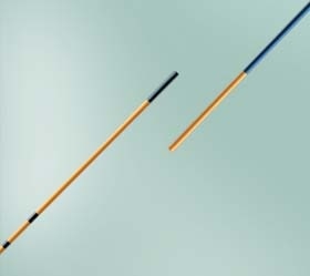
BD Solo™ Plus Guidewire
The BD Solo™ Plus Guidewire is designed for access to the ureter and maneuverability around stone. With its radiopaque tip, the Solo™ Plus is visible and detectible under fluoroscopic view. The BD Solo™ Plus is also 41% stiffer2 than the leading competition.
BD offers a full line of guidewires including the BD Solo™ Flex and Hydro.
Dilation
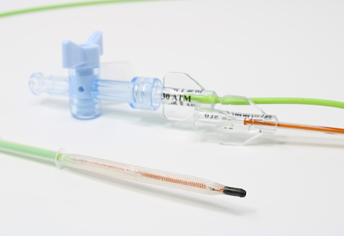
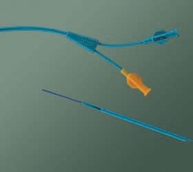
The Proxis™ Ureteral Access Sheath & X-Force™ Balloon Dilation Catheter
The X-Force™ line of balloon dilation catheters are comprised of a patented multilayer balloon with a rated burst pressure of 30ATM. Utilized in the management of urolithiasis and ureteric strictures, the X-Force™ boasts a radiopaque tungsten tip to aid in radiographic visualization.
The Proxis™ Ureteral Access Sheath combines hydrophilic coating, a smooth dilator-sheath transition, and a large hub to aid in access and introduction of instruments.

Visualization
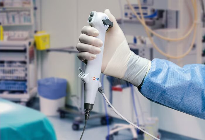
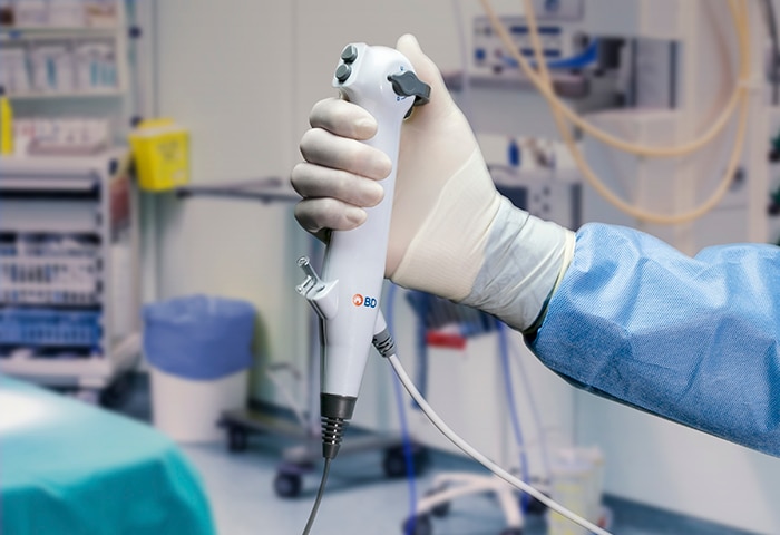
BD Aptra™ Ureteroscope
The BD Aptra™ Single-Use Digital Flexible Ureteroscope offers image quality enhanced with the light source inside the tip of the scope, tapered 7.4 Fr. distal tip, and maximum deflection designed to reach stones in the lower pole.
Fragmentation
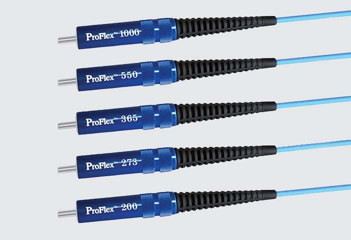
ProFlex™ Laser Fibers
The ProFlex™ Laser Fiber is a fiber optic laser delivery system consisting of a patented Pulsarill HPC high power connector on a silica core, double clad fiber.
Removal
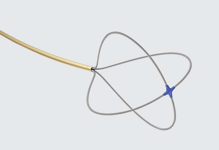
SkyLite™ Tipless Nitinol Basket
Constructed of Nitinol, the SkyLite™ Basket offers a kink-resistant design to aid in stone extraction and is designed to perform in a fully deflected flexible ureteroscope.

Drainage
Inlay Optima™ Ureteral Stent
Stent designed for ease of insertion, navigation around obstacles, and minimized migration. pHreeCoat provides a hydrophobic coating with smooth surface and encrustation resistant properties.
-
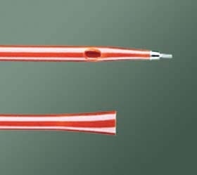
BD ureteral catheter choices are all carefully crafted to help you achieve two clinical objectives: efficient access to sometimes difficult and delicate anatomy and a minimal risk of trauma to tissue.
-
The dual lumen catheter provides a .038" diameter guidewire lumen and a .050" diameter injection lumen.
-
Ureteral catheters can be used for a number of functions such as maintaining drainage, infusion of contrast media and ureteral dilation.
-
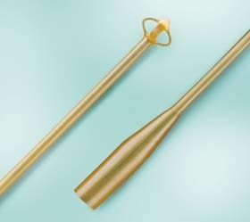
BD offers a wide range of latex drains. These include Pezzers, Malecots, and Angle Drains.
-
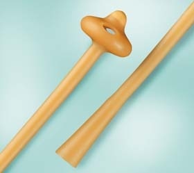
BD offers a wide range of latex drains. These include Pezzers, Malecots, and Angle Drains.
-
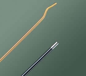
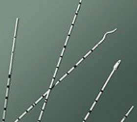
BD offers a wide array of filiforms and followers. Filiforms are used to get past difficult strictures whereas followers are used for dilation and drainage.
-
BD offers a wide array of filiforms and followers.
1. Data based on IPSOS report for BD.
2. 0.035" guidewires tested. Boston Scientific Sensor® Dual-Flex tested. Bench data on file with BD. In-vitro data may not correlate to clinical performance.
Intended use
BD Aptra™ Digital Endoscope System is intended to be used by physicians to access, visualize, and perform procedures in the urinary tract and the kidney. The instrument enables delivery and use of accessories such as biopsy forceps, laser fibers, graspers and retrieval baskets at a surgical site.
Contraindications
Diagnostic or therapeutic ureteroscopy is contraindicated in people with an untreated urinary tract infection.
Other contraindications to therapeutic ureteroscopy (e.g. lithotripsy, endopyelotomy, tumor therapy) are more numerous and can mirror those associated with the corresponding open surgical interventions. Patients on anticoagulants or with coagulopathies should be managed appropriately.
Warnings
• Do not use electromedical energy sources in the presence of flammable detergents, anesthetics, nitrous oxide (N2O), or oxygen.
• Consult the user manuals of all electromedical energy sources used with endoscopic instruments for appropriate instruments, warnings and cautions prior to use. Such sources of energy include electrical, electrohydraulic, electrosurgical, heat hydraulic, laser, light, pressure, sound, ultrasound and vacuum.
• Do not insert or advance the ureteroscope unless there is a clear live endoscopic view of the lumen through which the scope is being advanced (or confirm with visualization by other imaging modalities).
• During the procedure, if the live endoscopic image is lost, do not advance or insert the ureteroscope and do not insert, advance or actuate accessories.
• Do not use excessive force while advancing or withdrawing the scope. If resistance is felt during advancement or withdrawal of the scope, investigate the source of resistance and/or take remedial action if necessary.
• Do not force the distal tip of the ureteroscope against the sidewall of the ureter or renal pelvis.
• Do not use excessive force when advancing or withdrawing an accessory within the ureteroscope.
• When inserting or using accessories, maintain continuous visualization of the distal tip. Ensure that the distance between the distal tip of the ureteroscope and the object in view is greater than the ureteroscope’s minimum visible distance. Failure to do so may result in the accessories causing patient injury.
• Do not withdraw a laser fiber back into the ureteroscope while the laser is firing. Doing so may cause patient injury and/or scope damage.
• Do not look directly into the light emitted from the ureteroscope.
• Verify ground isolation when setting up and using accessories from different manufacturers prior to procedure.
• Do not open the handle of the ureteroscope.
• The ureteroscope is a single-use device and there are no serviceable parts. Do not repair damaged or non-operating ureteroscopes. Do not use the ureteroscope if damage is discovered or suspected.
• Do not excessively bend the flexible shaft or the articulating section of the ureteroscope.
• If damage to the ureteroscope occurs or it stops functioning during a procedure, stop using the ureteroscope immediately. See troubleshooting section for more information. Continue the procedure with a new ureteroscope, as appropriate.